RACP Divisional Written Exam · FRACP
Cardiology for the written exam
Everything Physify has for Cardiology: short lessons written to Australian guidance, exam-style questions with a worked explanation for each, and Bedside, our AI tutor, under every one. Try a question below, no sign-up.
- courses
- 35courses
- lessons
- 157lessons
- exam-style questions
- 1,094exam-style questions
- figures in the lessons
- 32figures in the lessons
Try one now · no sign-up
Cardiology
Pick an answer to see the explanation
What's covered
35 courses, each a set of lessons with a quiz after them. The bar shows how many questions each has. Open a course to see its lesson outline.
- Acute Coronary Syndromes4 lessons · 39 Qs
- Adult Congenital Heart Disease5 lessons · 32 Qs
- Aortic Disease4 lessons · 32 Qs
- Aortic Valve Disease4 lessons · 31 Qs
- Atrial Fibrillation and Flutter5 lessons · 39 Qs
- Australian Guidance Versus Overseas: Cardiovascular, Renal, Endocrine, Respiratory and Neurology4 lessons · 30 Qs
- Bradycardia and Conduction Disease4 lessons · 33 Qs
- Cardiac Amyloidosis and the Heart in Systemic Disease4 lessons · 27 Qs
- Cardiac Arrest, Peri-Arrest Algorithms and Post-Resuscitation Care5 lessons · 29 Qs
- Cardiac Devices: Pacemakers, ICDs and CRT4 lessons · 27 Qs
- Cardiac Investigations3 lessons · 25 Qs
- Cardiology in Special Populations and Perioperative Risk10 lessons · 25 Qs
- Cardiomyopathies4 lessons · 33 Qs
- Cardiovascular Pharmacology5 lessons · 31 Qs
- Cardiovascular Physiology5 lessons · 30 Qs
- Cardiovascular Risk & Primary Prevention5 lessons · 31 Qs
- ECG Interpretation4 lessons · 36 Qs
- Heart Failure4 lessons · 36 Qs
- Hypertension6 lessons · 46 Qs
- Infective Endocarditis and Prosthetic Valves5 lessons · 36 Qs
- Landmark Trials 2016 to 2026: Cardiology5 lessons · 27 Qs
- Landmark Trials: Cardiology and Nephrology5 lessons · 30 Qs
- Mitral, Tricuspid and Pulmonary Valve Disease4 lessons · 30 Qs
- Myocarditis3 lessons · 20 Qs
- Pericardial Disease4 lessons · 32 Qs
- Peripheral Arterial Disease3 lessons · 25 Qs
- Pulmonary Hypertension4 lessons · 31 Qs
- Reading the 12-Lead ECG: Real Tracings7 lessons · 53 Qs
- Rheumatic Fever and Rheumatic Heart Disease3 lessons · 20 Qs
- Shock, Haemodynamics and Vasopressors5 lessons · 30 Qs
- Stable Ischaemic Heart Disease3 lessons · 31 Qs
- Supraventricular Tachycardias4 lessons · 27 Qs
- Syncope and Sudden Cardiac Death4 lessons · 27 Qs
- The Endothelium and Lipoproteins5 lessons · 30 Qs
- Ventricular Arrhythmias and Channelopathies4 lessons · 33 Qs
From the lessons
32 figures sit in the Cardiology lessons: ECGs, diagrams, films and clinical images, each with what to look for. Here are 4. Tap one to enlarge it.
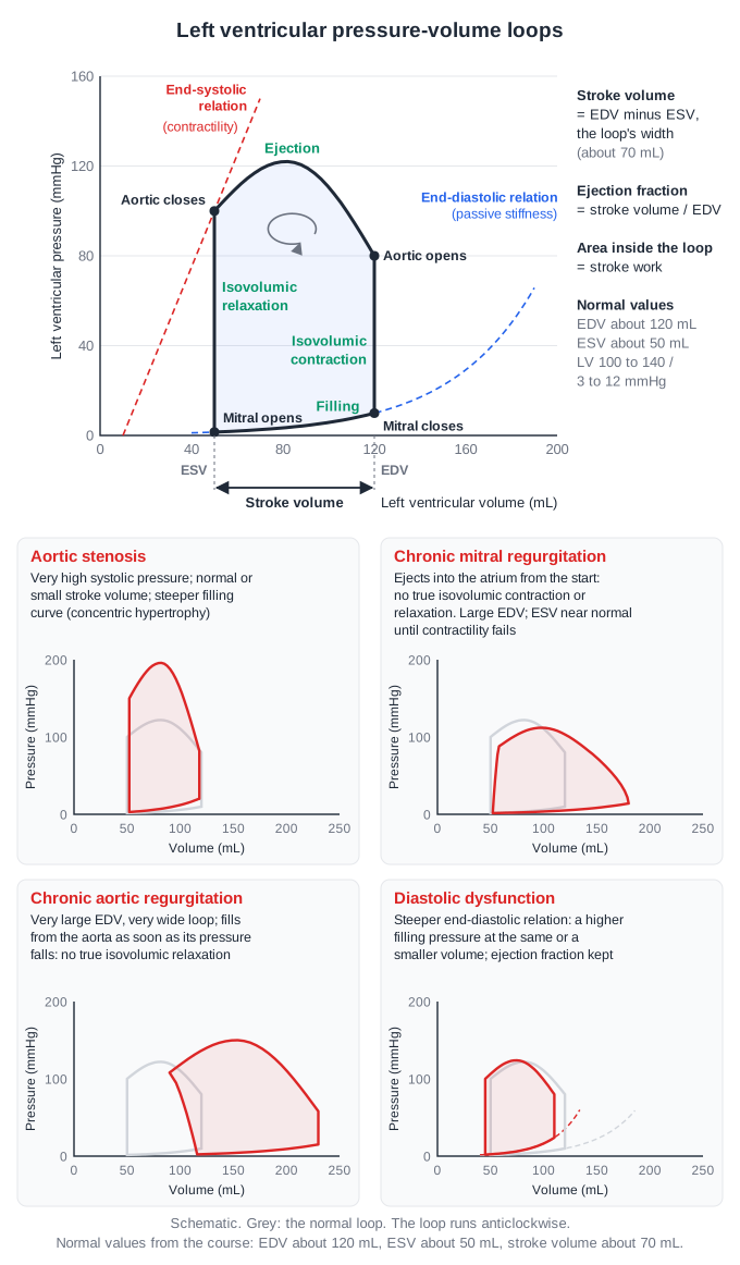
From the lesson “Ventricular Mechanics, Coronary Flow and Oxygen Transport” in Cardiovascular Physiology
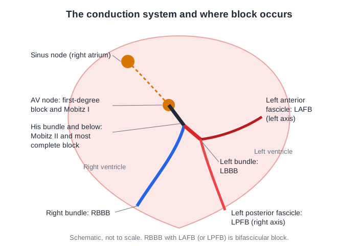
From the lesson “Bundle Branch and Fascicular Block” in Bradycardia and Conduction Disease
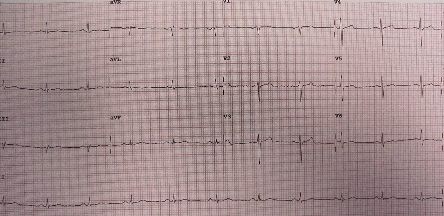
From the lesson “Mimics and Hidden Signs: Repolarisation, Electrolytes, Ischaemia and Inherited Disease” in Reading the 12-Lead ECG: Real Tracings
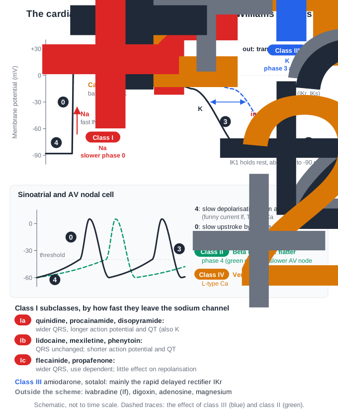
From the lesson “Antiarrhythmics and Rate-Controlling Agents” in Cardiovascular Pharmacology
Free for Cardiology
Cardiology in the clinical exam
25 interactive short cases and 6 long cases for the RACP Divisional Clinical Exam: you examine, present and are marked on the five RACP domains.
See the clinical exam →Cardiology on the blog
All posts →Brugada Syndrome: Trials of Note for the RACP Exam
Brugada syndrome for the RACP exam: the type 1 ECG, the 2022 ESC rule on drug-induced patterns, ICD or not, fever and drugs to avoid, and ablation (BRAVE).
Rheumatic Heart Disease: Trials of Note for the RACP Exam
Rheumatic heart disease for the RACP exam: Edition 3.4 of the Australian guideline, ARF criteria by risk group, benzathine prophylaxis, GOAL and INVICTUS.
Sick Sinus Syndrome: Key Points for the RACP Exam
Sick sinus syndrome for the RACP exam: its forms, reversible causes, when to pace, why dual-chamber pacing wins (DANPACE), and AF in tachy-brady syndrome.
Torsades de Pointes: Key Points for the RACP Exam
Torsades de pointes for the RACP exam: recognising it, the drugs and risk factors, the ANZCOR magnesium regimen, pacing and isoprenaline, congenital long QT.
Aortic Regurgitation: Trials of Note for the RACP Exam
Aortic regurgitation for the RACP exam in 2026: when to operate, the Australian rheumatic guideline, indexed LV thresholds and dedicated TAVI valves (ALIGN-AR).
Aortic Stenosis: Trials of Note for the RACP Exam
Aortic stenosis for the RACP exam in 2026: severity thresholds, TAVI or surgery by age, early TAVI for asymptomatic disease, and PROGRESS in moderate stenosis.
Open all of Cardiology
A membership opens every lesson and all 1,094 Cardiology questions, with the rest of the bank (11,500+ questions across 17 specialties), 15 timed mock exams, smart review and a daily plan to your exam.
The trial needs a card; cancel any time before it ends and you pay nothing. The free account needs no card.