RACP Divisional Written Exam · FRACP
Renal for the written exam
Everything Physify has for Renal: short lessons written to Australian guidance, exam-style questions with a worked explanation for each, and Bedside, our AI tutor, under every one. Try a question below, no sign-up.
- courses
- 30courses
- lessons
- 163lessons
- exam-style questions
- 801exam-style questions
- figures in the lessons
- 21figures in the lessons
Try one now · no sign-up
Renal
Pick an answer to see the explanation
What's covered
30 courses, each a set of lessons with a quiz after them. The bar shows how many questions each has. Open a course to see its lesson outline.
- Acid-Base Disorders5 lessons · 30 Qs
- Acute Interstitial Nephritis4 lessons · 20 Qs
- Acute Kidney Injury5 lessons · 51 Qs
- Australian Guidance Versus Overseas: Cardiovascular, Renal, Endocrine, Respiratory and Neurology4 lessons · 30 Qs
- Cardiorenal Syndrome4 lessons · 25 Qs
- Chronic Kidney Disease6 lessons · 20 Qs
- Diabetic Nephropathy4 lessons · 25 Qs
- Electrolyte Abnormalities12 lessons · 30 Qs
- Fluid Therapy and Volume Assessment5 lessons · 29 Qs
- Genetic Kidney Diseases9 lessons · 31 Qs
- Hypertension6 lessons · 46 Qs
- Inherited Tubular and Metabolic Kidney Disease6 lessons · 20 Qs
- Landmark Trials 2016 to 2026: Nephrology5 lessons · 26 Qs
- Landmark Trials: Cardiology and Nephrology5 lessons · 30 Qs
- Milk Alkali Syndrome3 lessons · 8 Qs
- Monoclonal Gammopathy of Renal Significance4 lessons · 25 Qs
- Myeloma Kidney4 lessons · 25 Qs
- Nephrotic vs. Nephritic Syndromes10 lessons · 62 Qs
- Reflux Nephropathy & Urinary Tract Infection5 lessons · 20 Qs
- Renal Amyloidosis5 lessons · 25 Qs
- Renal Calculi & Nephrocalcinosis5 lessons · 20 Qs
- Renal Physiology5 lessons · 20 Qs
- Renal Replacement Therapy6 lessons · 21 Qs
- Renal Sarcoidosis4 lessons · 9 Qs
- Renal Transplantation6 lessons · 20 Qs
- Renal Tubular Acidosis4 lessons · 25 Qs
- Renovascular Disease6 lessons · 21 Qs
- Thrombotic Microangiopathies6 lessons · 31 Qs
- Toxic Nephropathy5 lessons · 25 Qs
- Urinary Tract Obstruction, Post-Obstructive Diuresis & Renal Tumours5 lessons · 31 Qs
From the lessons
21 figures sit in the Renal lessons: ECGs, diagrams, films and clinical images, each with what to look for. Here are 4. Tap one to enlarge it.
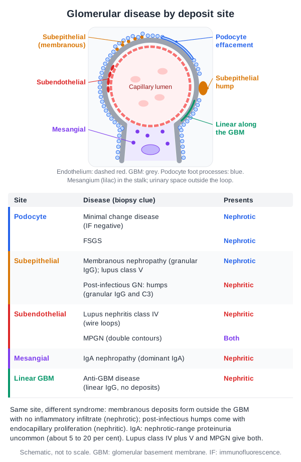
From the lesson “Approach, Classification and Biopsy” in Nephrotic vs. Nephritic Syndromes
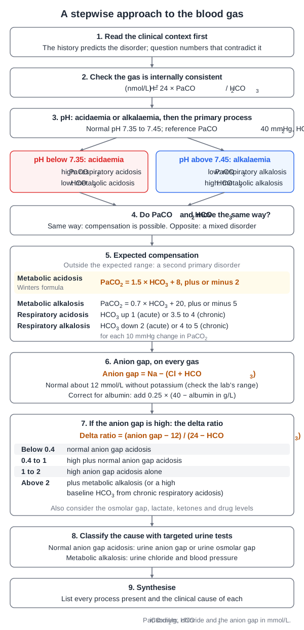
From the lesson “Physiology and a Stepwise Approach to the Blood Gas” in Acid-Base Disorders
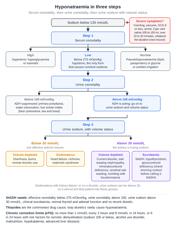
From the lesson “Hyponatraemia: Classification and Diagnosis” in Electrolyte Abnormalities
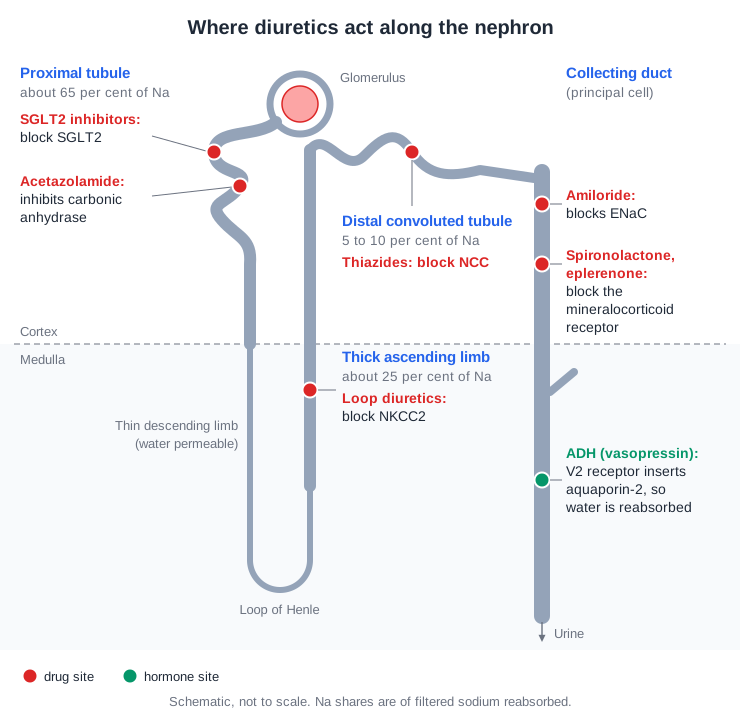
From the lesson “Tubular Transport by Segment” in Renal Physiology
Free for Renal
Renal in the clinical exam
7 interactive short cases and 4 long cases for the RACP Divisional Clinical Exam: you examine, present and are marked on the five RACP domains.
See the clinical exam →Renal on the blog
All posts →Acute Interstitial Nephritis: Recent Advances
Acute interstitial nephritis for the RACP exam: drug causes, checkpoint inhibitor AKI and ADQI 2026, early steroids, rechallenge, and the urinary CXCL9 test.
Chronic Kidney Disease and Reflux Nephropathy: Current and Emerging Therapy
CKD and reflux nephropathy for the RACP exam: the Kidney Health Australia handbook, ACE inhibitor or ARB plus SGLT2 inhibitor, PBS status, and the RIVUR trial.
Electrolyte Disorders: Key Therapies for the RACP Exam
Electrolytes for the RACP exam: hypertonic saline and SALSA, tolvaptan limits, patiromer and SZC with PBS status, and denosumab for refractory hypercalcaemia.
Renal Calculi (Kidney Stones): Advances and Management
Kidney stones for the RACP exam: the 2026 CARI guideline, fluid, sodium and calcium targets, why NOSTONE demoted thiazides, potassium citrate and lumasiran.
Renal Replacement Therapy: Key Evidence and Evolving Practice
Dialysis timing and dose for the RACP exam: STARRT-AKI, AKIKI 2, RENAL and IDEAL, the KHA-CARI adaptation of KDIGO, and what ANZDATA 2025 shows about care.
Renal Sarcoidosis: Evidence-Based Management
Renal sarcoidosis for the RACP exam: granulomatous interstitial nephritis, calcitriol-driven hypercalcaemia, steroid response in the cohorts, and relapse.
Open all of Renal
A membership opens every lesson and all 801 Renal questions, with the rest of the bank (11,500+ questions across 17 specialties), 15 timed mock exams, smart review and a daily plan to your exam.
The trial needs a card; cancel any time before it ends and you pay nothing. The free account needs no card.