RACP Divisional Written Exam · FRACP
Rheumatology/Immunology for the written exam
Everything Physify has for Rheumatology/Immunology: short lessons written to Australian guidance, exam-style questions with a worked explanation for each, and Bedside, our AI tutor, under every one. Try a question below, no sign-up.
- courses
- 37courses
- lessons
- 130lessons
- exam-style questions
- 947exam-style questions
- figures in the lessons
- 29figures in the lessons
Try one now · no sign-up
Rheumatology/Immunology
Pick an answer to see the explanation
What's covered
37 courses, each a set of lessons with a quiz after them. The bar shows how many questions each has. Open a course to see its lesson outline.
- Allergy, Anaphylaxis & Angio-oedema4 lessons · 23 Qs
- Antibody & Combined Immunodeficiency3 lessons · 30 Qs
- Approach to the Patient with Joint Pain3 lessons · 20 Qs
- Australian Guidance Versus Overseas: Infection, Haematology, Gastroenterology, Rheumatology and Oncology5 lessons · 30 Qs
- Autoantibodies & Immunological Testing3 lessons · 20 Qs
- Autoimmune Cytopenias3 lessons · 30 Qs
- Autoinflammatory & Periodic Fever Syndromes3 lessons · 30 Qs
- Bone & Joint Infection3 lessons · 31 Qs
- Crystalline Arthropathies4 lessons · 32 Qs
- Food Allergy5 lessons · 27 Qs
- Haemophagocytic Syndromes & Cytokine Release3 lessons · 20 Qs
- Heritable Disorders of Connective Tissue3 lessons · 20 Qs
- Human Immunodeficiency Virus (HIV) & AIDS3 lessons · 21 Qs
- IgG4 Related Disease3 lessons · 20 Qs
- Immunosuppressants & Biologics3 lessons · 30 Qs
- Inflammatory Myopathies & Polymyalgia Rheumatica3 lessons · 30 Qs
- Inflammatory/Immunological Eye Disorders3 lessons · 20 Qs
- Landmark Trials 2016 to 2026: Rheumatology and Immunology5 lessons · 27 Qs
- Landmark Trials: Neurology, Rheumatology, Geriatrics and Acute Care5 lessons · 29 Qs
- Large Vessel Vasculitides3 lessons · 20 Qs
- Malignancy & Tumour-like Joint Lesions3 lessons · 20 Qs
- Medium & Variable Vessel Vasculitis3 lessons · 30 Qs
- Metabolic, Endocrine & Haematological Arthropathies3 lessons · 30 Qs
- Osteoarthritis & Regional Musculoskeletal Disorders3 lessons · 20 Qs
- Phagocyte, Regulatory & Lymphoproliferative Defects3 lessons · 20 Qs
- Principles of Immunology5 lessons · 31 Qs
- Rare Rheumatological Syndromes3 lessons · 20 Qs
- Rheumatic Disease in Pregnancy3 lessons · 20 Qs
- Rheumatoid Arthritis4 lessons · 31 Qs
- Sarcoidosis5 lessons · 21 Qs
- Scleroderma3 lessons · 32 Qs
- Seronegative Spondyloarthropathies4 lessons · 30 Qs
- Sjogren's Syndrome3 lessons · 20 Qs
- Small-Vessel Vasculitides3 lessons · 31 Qs
- Systemic Lupus Erythematosus4 lessons · 30 Qs
- Transplant Immunology and HLA5 lessons · 31 Qs
- Viral & Post-infectious Arthritis3 lessons · 20 Qs
From the lessons
29 figures sit in the Rheumatology/Immunology lessons: ECGs, diagrams, films and clinical images, each with what to look for. Here are 4. Tap one to enlarge it.
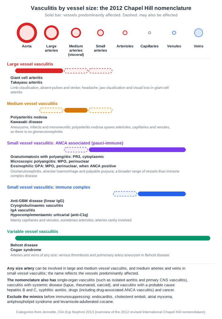
From the lesson “ANCA Testing & The Clinical Syndromes” in Small-Vessel Vasculitides
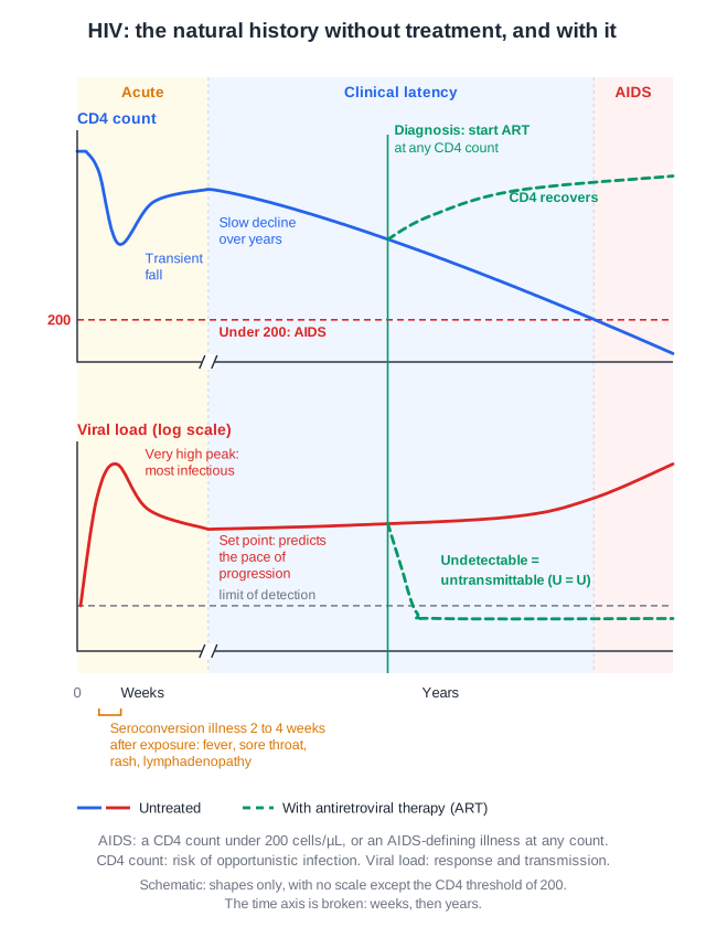
From the lesson “Testing, Staging & Antiretroviral Therapy” in Human Immunodeficiency Virus (HIV) & AIDS
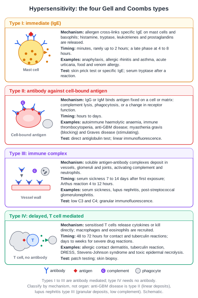
From the lesson “Tolerance, Autoimmunity, Immune Checkpoints and Hypersensitivity” in Principles of Immunology
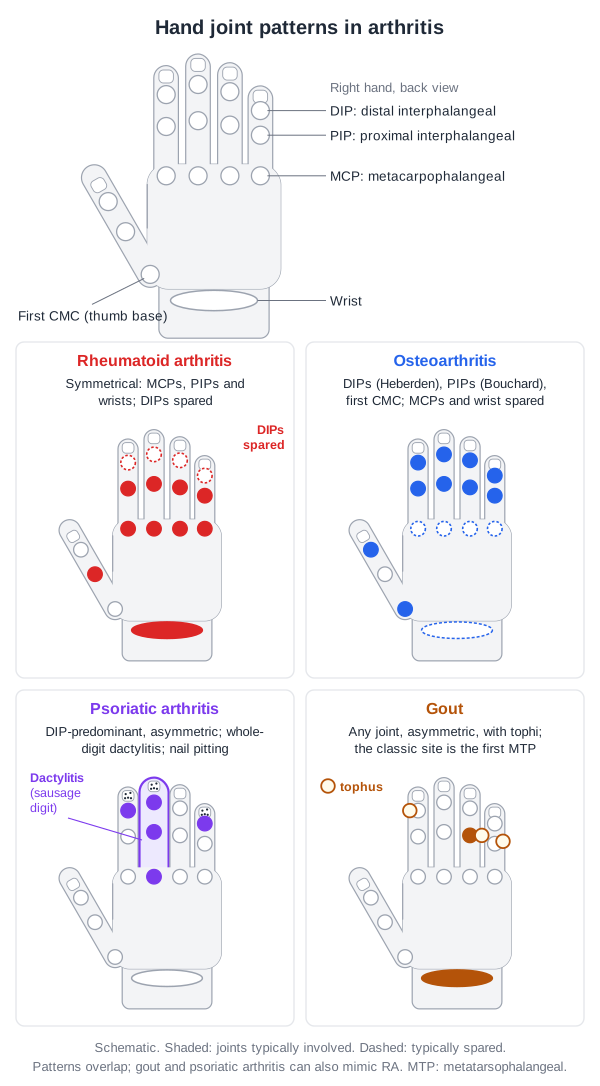
From the lesson “Articular Patterns & The Acute Monoarthritis” in Approach to the Patient with Joint Pain
Free for Rheumatology/Immunology
Rheumatology/Immunology in the clinical exam
14 interactive short cases and 5 long cases for the RACP Divisional Clinical Exam: you examine, present and are marked on the five RACP domains.
See the clinical exam →Rheumatology/Immunology on the blog
All posts →Behçet's Disease: Current and Emerging Therapies
Behçet's disease for the RACP exam: treatment by organ under the 2025 EULAR update, colchicine first, apremilast (RELIEF), early anti-TNF and the PBS position.
Crystalline Arthropathies (Gout and CPPD): Advances and Management
Gout and CPPD for the RACP exam: treat urate to below 0.36 mmol/L, allopurinol first, febuxostat after CARES and FAST, pegloticase with methotrexate, CPPD.
Ehlers-Danlos Syndrome: Modern and Emerging Therapies
Ehlers-Danlos syndrome for the RACP exam: the 13 subtypes, why vascular EDS matters, celiprolol (BBEST), irbesartan added in 2025, and Australian funding.
Emerging Therapies for Polymyalgia Rheumatica and Inflammatory Myositis: RACP Exam Notes
PMR and myositis for the RACP exam: 2025 EULAR prednisolone doses, sarilumab (SAPHYR) and secukinumab (REPLENISH) for relapse, IVIG in dermatomyositis, funding.
Emerging Therapies for Sjögren's Disease: RACP Exam Notes
Sjögren's disease for the RACP exam: EULAR 2019 treatment by symptom and organ, the modest NEPTUNUS ianalumab results, CD40 and FcRn trials, and access.
Emerging Therapies for Systemic Lupus Erythematosus: RACP Exam Notes
SLE for the RACP exam: EULAR 2023 hydroxychloroquine and steroid targets, anifrolumab, belimumab and obinutuzumab in nephritis, new trials and PBS status.
Open all of Rheumatology/Immunology
A membership opens every lesson and all 947 Rheumatology/Immunology questions, with the rest of the bank (11,500+ questions across 17 specialties), 15 timed mock exams, smart review and a daily plan to your exam.
The trial needs a card; cancel any time before it ends and you pay nothing. The free account needs no card.