RACP Divisional Written Exam · FRACP

Medical Sciences for the written exam

Everything Physify has for Medical Sciences: short lessons written to Australian guidance, exam-style questions with a worked explanation for each, and Bedside, our AI tutor, under every one. Try a question below, no sign-up.

courses
44courses
lessons
227lessons
exam-style questions
1,187exam-style questions
figures in the lessons
36figures in the lessons

Try one now · no sign-up

Medical Sciences

A 74 year old man with atrial fibrillation starts digoxin 125 micrograms daily with no loading dose. His creatinine is 118 micromol/L, eGFR 44 mL/min/1.73 m2, potassium 4.3 mmol/L and weight 71 kg, and at this level of renal function his digoxin half-life is about 60 hours. When will his concentration be at steady state?

Pick an answer to see the explanation

What's covered

44 courses, each a set of lessons with a quiz after them. The bar shows how many questions each has. Open a course to see its lesson outline.

From the lessons

36 figures sit in the Medical Sciences lessons: ECGs, diagrams, films and clinical images, each with what to look for. Here are 4. Tap one to enlarge it.
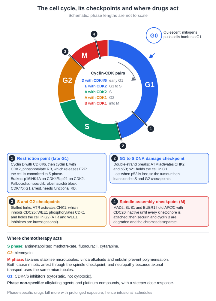
Schematic of the cell cycle as a ring (phase lengths not to scale): G1, S, G2 and M, with quiescent G0 outside it and mitogens pushing cells back into G1. The cyclin-CDK pairs are listed in the centre: cyclin D with CDK4/6 in early G1, cyclin E with CDK2 at G1 to S, cyclin A with CDK2 in S, cyclin A with CDK1 in G2 and cyclin B with CDK1 into M. Four numbered checkpoints are marked. 1, the restriction point in late G1: cyclin D with CDK4/6 and then cyclin E with CDK2 phosphorylate RB, which releases E2F and commits the cell to S phase; p16INK4A brakes CDK4/6 and p21 brakes CDK2; palbociclib, ribociclib and abemaciclib block CDK4/6 and cause G1 arrest, which needs functional RB. 2, the G1 to S DNA damage checkpoint: ATM activates CHK2 and p53, and p21 holds the cell in G1; it is lost with p53, so the tumour leans on the S and G2 checkpoints. 3, the S and G2 checkpoints: ATR activates CHK1, which inhibits CDC25, and WEE1 phosphorylates CDK1 to hold the cell in G2 (ATR and WEE1 inhibitors are investigational). 4, the spindle assembly checkpoint in M: MAD2, BUB1 and BUBR1 hold APC/C with CDC20 inactive until every kinetochore is attached. Below, where chemotherapy acts: antimetabolites (methotrexate, fluorouracil, cytarabine) in S phase; bleomycin in G2; taxanes, which stabilise microtubules, and vinca alkaloids and eribulin, which prevent polymerisation, in M phase, causing mitotic arrest and neuropathy; CDK4/6 inhibitors in G1, cytostatic rather than cytotoxic; and alkylating agents and platinum compounds as phase non-specific, with a steeper dose-response. Phase-specific drugs kill more with prolonged exposure, hence infusional schedules.
Physify teaching diagram (schematic). Tap the image to zoom.

From the lesson “The Cell Cycle, p53, Checkpoints and DNA Repair” in Cell, Molecular and Cancer Biology
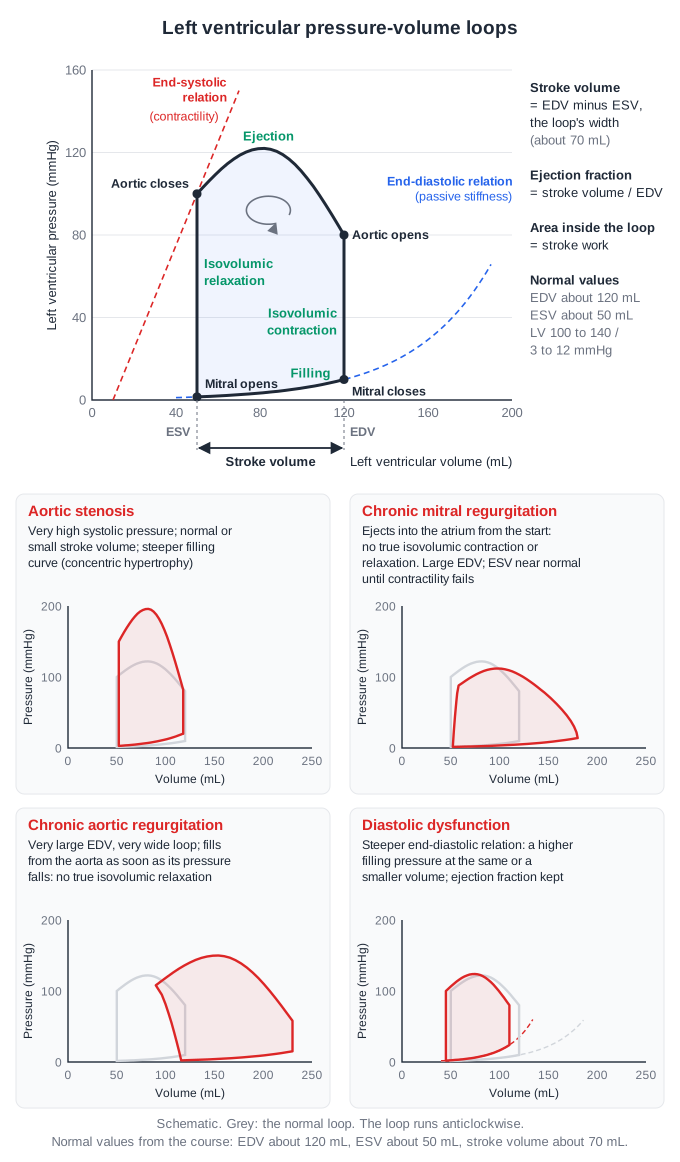
Schematic left ventricular pressure-volume loop, volume on the x axis and pressure on the y axis, running anticlockwise. Mitral closure at end-diastole (about 120 mL) starts isovolumic contraction up the right side; the aortic valve opens at about 80 mmHg; ejection runs along the top, peaking near 120 mmHg; the aortic valve closes at end-systole (about 50 mL, about 100 mmHg); isovolumic relaxation falls down the left side; the mitral valve opens and filling runs along the bottom. The width of the loop is the stroke volume, about 70 mL; ejection fraction is stroke volume divided by end-diastolic volume and the area inside the loop is stroke work. A dashed straight end-systolic pressure-volume relation (contractility) touches the end-systolic corner, and a dashed exponential end-diastolic relation (passive stiffness) runs along the filling limb. Four smaller panels compare abnormal loops with the normal one: aortic stenosis, a tall loop with very high systolic pressure, a normal or small stroke volume and a steeper filling curve; chronic mitral regurgitation, a large end-diastolic volume and ejection into the atrium from the start, so no true isovolumic contraction or relaxation; chronic aortic regurgitation, a very large end-diastolic volume and very wide loop with filling from the aorta during relaxation, so no true isovolumic relaxation; and diastolic dysfunction, a steeper end-diastolic relation giving a higher filling pressure at the same or a smaller volume.
Physify teaching diagram (schematic). Tap the image to zoom.

From the lesson “Ventricular Mechanics, Coronary Flow and Oxygen Transport” in Cardiovascular Physiology
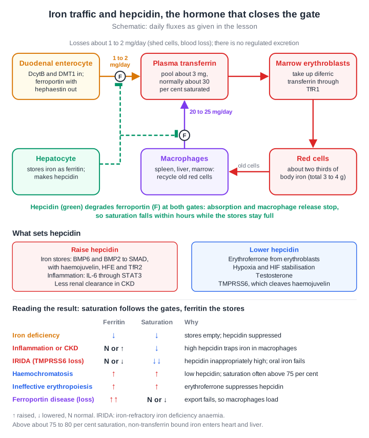
Schematic of iron traffic. The duodenal enterocyte takes iron in through duodenal cytochrome b and DMT1 and exports it through ferroportin with hephaestin, about 1 to 2 mg a day, into plasma transferrin, a pool of about 3 mg that is normally about 30 per cent saturated. Marrow erythroblasts take up diferric transferrin through TfR1 and make red cells, which hold about two thirds of the 3 to 4 g of body iron. Macrophages recycle old red cells and return 20 to 25 mg a day to plasma through ferroportin. Losses are about 1 to 2 mg a day from shed cells and blood loss; there is no regulated excretion. The hepatocyte stores iron as ferritin and makes hepcidin, which degrades ferroportin at both gates, the enterocyte and the macrophage, so absorption and macrophage release stop and saturation falls within hours while the stores stay full. Hepcidin is raised by iron stores through BMP6 and BMP2 to SMAD, with haemojuvelin, HFE and transferrin receptor 2, by inflammation through IL-6 and STAT3, and by reduced renal clearance in CKD; it is lowered by erythroferrone, hypoxia and HIF stabilisation, testosterone and TMPRSS6, which cleaves haemojuvelin. A table reads the results: iron deficiency, ferritin and saturation low; inflammation or CKD, ferritin normal or high with low saturation; TMPRSS6 loss (iron-refractory iron deficiency anaemia, IRIDA), ferritin normal or low with very low saturation; haemochromatosis and ineffective erythropoiesis, both high, saturation often above 75 per cent in haemochromatosis; and loss-of-function ferroportin disease, ferritin very high with normal or low saturation. Above about 75 to 80 per cent saturation, non-transferrin bound iron enters heart and liver.
Physify teaching diagram (schematic). Tap the image to zoom.

From the lesson “Iron: Absorption, Transport and the Hepcidin-Ferroportin Axis” in Oxygen Sensing, Iron and Haem
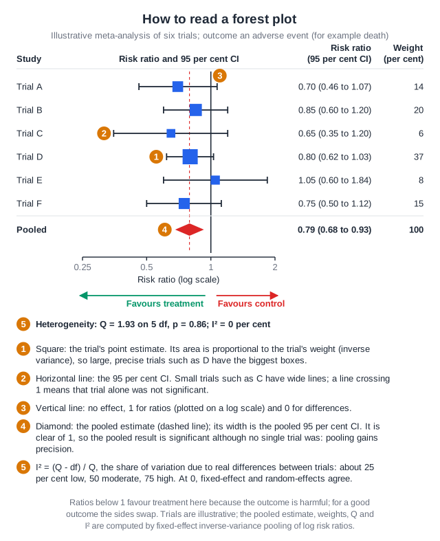
An illustrative forest plot of six trials with a harmful outcome such as death, risk ratios on a log scale from 0.25 to 2. Each trial is a blue square at its point estimate, with area proportional to its inverse-variance weight, on a horizontal line showing its 95 per cent confidence interval; trial D, the largest, carries 37 per cent of the weight and trial C, the smallest, 6 per cent with the widest interval. Every trial's interval crosses the vertical line of no effect at 1. The pooled fixed-effect estimate is a red diamond at 0.79, its width the pooled interval of 0.68 to 0.93, which is clear of 1, so pooling gives a significant result although no single trial was. Ratios below 1 favour treatment and above 1 favour control. Heterogeneity is Q 1.93 on 5 degrees of freedom, p 0.86, with I squared 0 per cent, so fixed-effect and random-effects models agree. A key explains I squared as (Q minus df) / Q, about 25 per cent low, 50 moderate and 75 high, and that the null is 1 for ratios and 0 for differences.
Physify teaching diagram. Tap the image to zoom.

From the lesson “Meta-Analysis, Heterogeneity and Publication Bias” in Biostatistics & Evidence-Based Medicine

Free for Medical Sciences

Open all of Medical Sciences

A membership opens every lesson and all 1,187 Medical Sciences questions, with the rest of the bank (11,500+ questions across 17 specialties), 15 timed mock exams, smart review and a daily plan to your exam.

The trial needs a card; cancel any time before it ends and you pay nothing. The free account needs no card.

Other specialties

Physify is a study aid for exam preparation only. It is not medical advice and must not be used to guide the care of any patient. Terms