RACP Divisional Written Exam · FRACP
Haematology for the written exam
Everything Physify has for Haematology: short lessons written to Australian guidance, exam-style questions with a worked explanation for each, and Bedside, our AI tutor, under every one. Try a question below, no sign-up.
- courses
- 30courses
- lessons
- 110lessons
- exam-style questions
- 781exam-style questions
- figures in the lessons
- 24figures in the lessons
Try one now · no sign-up
Haematology
Pick an answer to see the explanation
What's covered
30 courses, each a set of lessons with a quiz after them. The bar shows how many questions each has. Open a course to see its lesson outline.
- Anaemia6 lessons · 21 Qs
- Aplastic Anaemia3 lessons · 20 Qs
- Approach to Abnormal Blood Counts & the Blood Film3 lessons · 32 Qs
- Australian Guidance Versus Overseas: Infection, Haematology, Gastroenterology, Rheumatology and Oncology5 lessons · 30 Qs
- Autoimmune Cytopenias3 lessons · 30 Qs
- Bleeding Disorders3 lessons · 31 Qs
- Cardiac Amyloidosis and the Heart in Systemic Disease4 lessons · 27 Qs
- Eosinophilia, Mastocytosis & Rare Myeloid Neoplasms3 lessons · 20 Qs
- Haematological Disorders in Pregnancy3 lessons · 30 Qs
- Haemoglobinopathies3 lessons · 21 Qs
- Haemolysis3 lessons · 21 Qs
- Haemostasis & Coagulation Testing4 lessons · 31 Qs
- Heparin-Induced Thrombocytopenia3 lessons · 20 Qs
- Inherited Bone Marrow Failure & Germline Predisposition3 lessons · 20 Qs
- Iron Overload & Chelation3 lessons · 20 Qs
- Landmark Trials 2016 to 2026: Haematology5 lessons · 28 Qs
- Landmark Trials: Haematology, Oncology and Infectious Diseases5 lessons · 30 Qs
- Leukaemia6 lessons · 33 Qs
- Lymphoma5 lessons · 30 Qs
- Microangiopathic Haemolytic Anaemia3 lessons · 20 Qs
- Myelodysplastic Syndromes3 lessons · 31 Qs
- Myeloproliferative Disorders3 lessons · 31 Qs
- Oxygen Sensing, Iron and Haem5 lessons · 31 Qs
- Plasma Cell Dyscrasias4 lessons · 32 Qs
- Splenomegaly, Hyposplenism & Apheresis3 lessons · 21 Qs
- Stem Cell Transplantation4 lessons · 20 Qs
- Thrombophilias3 lessons · 30 Qs
- Transfusion Medicine & Immunohaematology3 lessons · 30 Qs
- Tumour Lysis Syndrome3 lessons · 20 Qs
- Venous Thromboembolism & Anticoagulation3 lessons · 20 Qs
From the lessons
24 figures sit in the Haematology lessons: ECGs, diagrams, films and clinical images, each with what to look for. Here are 4. Tap one to enlarge it.
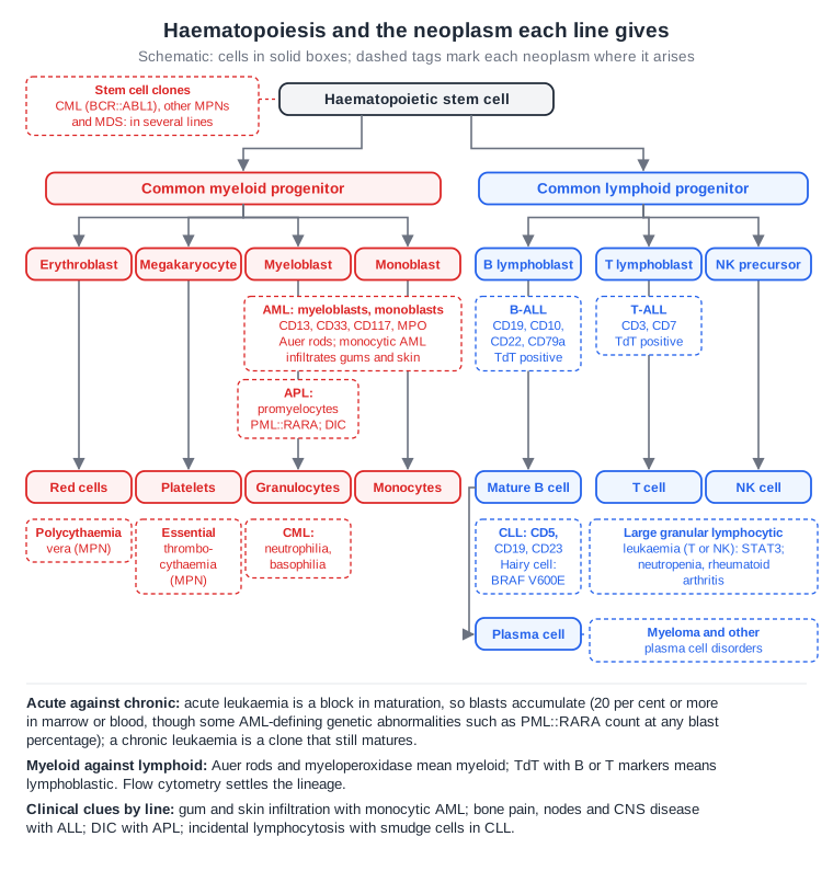
From the lesson “Diagnosis & Classification” in Leukaemia
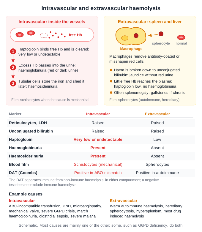
From the lesson “Recognising Haemolysis & Immune Causes” in Haemolysis
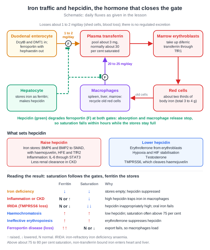
From the lesson “Iron: Absorption, Transport and the Hepcidin-Ferroportin Axis” in Oxygen Sensing, Iron and Haem
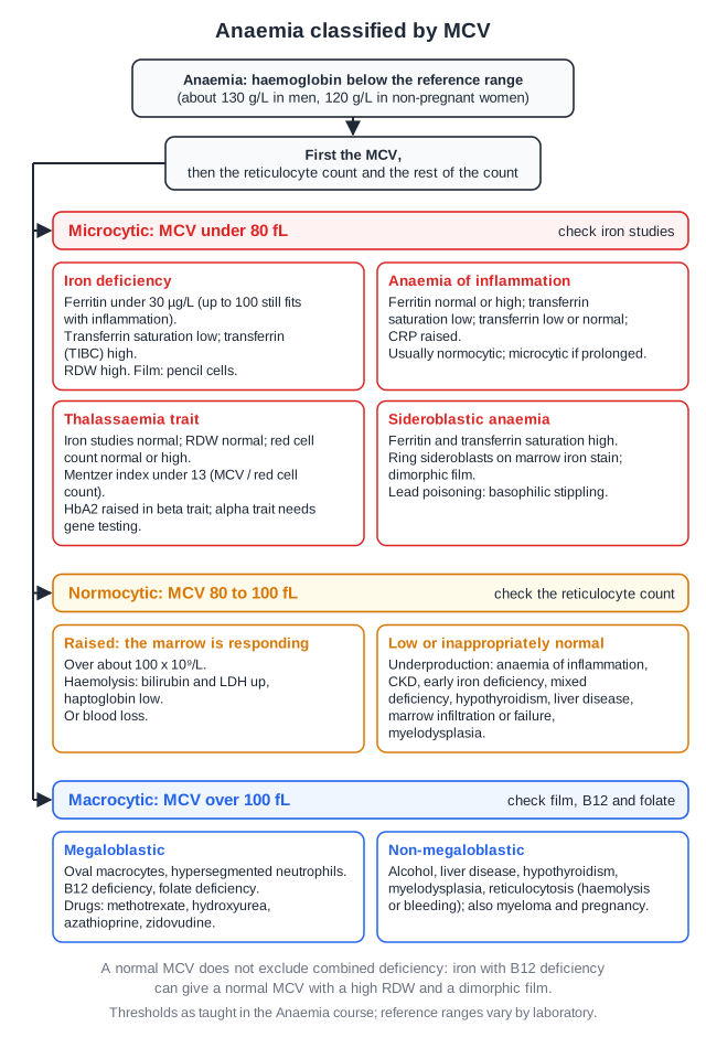
From the lesson “Approach to Anaemia” in Anaemia
Free for Haematology
Haematology in the clinical exam
10 interactive short cases and 3 long cases for the RACP Divisional Clinical Exam: you examine, present and are marked on the five RACP domains.
See the clinical exam →Haematology on the blog
All posts →Aplastic Anaemia: Trials of Note for the RACP Exam
Aplastic anaemia for the RACP exam: transplant versus horse ATG, ciclosporin and eltrombopag (RACE), the PBS first-line eltrombopag listing and eviQ protocols.
Complement-Mediated Thrombotic Microangiopathies: RACP Exam Notes
Complement-mediated TMA for the RACP exam: aHUS versus TTP, eculizumab and ravulizumab on the PBS, meningococcal vaccination, TGA-registered caplacizumab.
Disseminated Intravascular Coagulation: RACP Exam Notes
DIC for the RACP exam: ISTH scoring, treating the cause, when to give platelets, plasma and cryoprecipitate (Lifeblood, NBA), heparin, APL and SCARLET.
Haemolysis and Paroxysmal Nocturnal Haemoglobinuria: RACP Exam Notes
Haemolysis and PNH for the RACP exam: intravascular versus extravascular haemolysis, C5 and proximal inhibitors on the PBS, PEGASUS, APPLY-PNH and sutimlimab.
Heparin-Induced Thrombocytopenia: Key Points for the RACP Exam
HIT for the RACP exam: the 4Ts score, immunoassay then functional assay, danaparoid or fondaparinux at treatment dose (THANZ 2019), when DOACs fit, and VITT.
Leukaemia: Trials of Note for the RACP Exam
Leukaemia for the RACP exam: asciminib in CML, blinatumomab in MRD-negative ALL, venetoclax and FLT3 inhibitors in AML, with eviQ protocols and PBS status.
Open all of Haematology
A membership opens every lesson and all 781 Haematology questions, with the rest of the bank (11,500+ questions across 17 specialties), 15 timed mock exams, smart review and a daily plan to your exam.
The trial needs a card; cancel any time before it ends and you pay nothing. The free account needs no card.