RACP Divisional Written Exam · FRACP

Gastroenterology for the written exam

Everything Physify has for Gastroenterology: short lessons written to Australian guidance, exam-style questions with a worked explanation for each, and Bedside, our AI tutor, under every one. Try a question below, no sign-up.

courses
31courses
lessons
116lessons
exam-style questions
856exam-style questions
figures in the lessons
39figures in the lessons

Try one now · no sign-up

Gastroenterology

A 62 year old woman has acute liver failure with grade 2 encephalopathy. Lactate dehydrogenase is 3200 U/L, alkaline phosphatase 480 U/L and bilirubin 190 micromol/L, and the liver measures 18 cm with diffuse hypoechoic infiltration. Bone marrow examination shows a high grade B cell lymphoma. What does this mean for transplantation?

Pick an answer to see the explanation

What's covered

31 courses, each a set of lessons with a quiz after them. The bar shows how many questions each has. Open a course to see its lesson outline.

From the lessons

39 figures sit in the Gastroenterology lessons: ECGs, diagrams, films and clinical images, each with what to look for. Here are 4. Tap one to enlarge it.
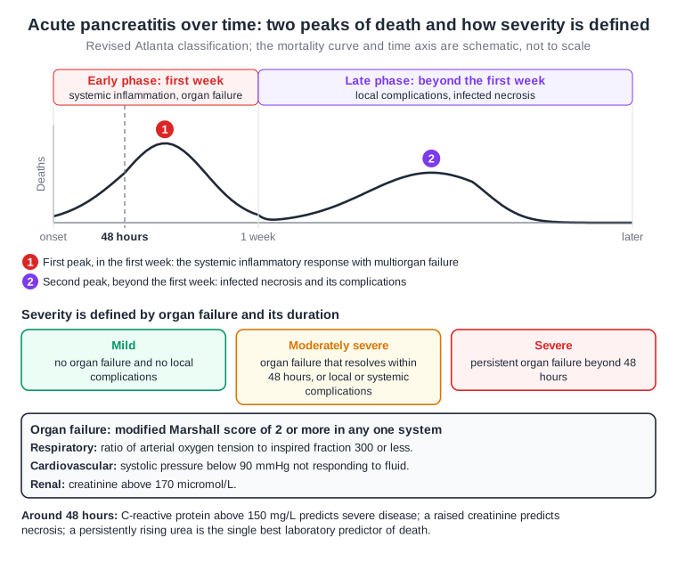
Timeline of acute pancreatitis from onset through 48 hours and 1 week; the curve and the time axis are schematic and not to scale. A curve of deaths shows two peaks: the first in the early phase (the first week) from the systemic inflammatory response with multiorgan failure, the second in the late phase, beyond the first week, from infected necrosis and its complications. Revised Atlanta severity: mild, no organ failure and no local complications; moderately severe, organ failure resolving within 48 hours or local or systemic complications; severe, organ failure persisting beyond 48 hours. Organ failure is a modified Marshall score of 2 or more in one system: arterial oxygen tension to inspired fraction ratio 300 or less, systolic pressure below 90 mmHg not responding to fluid, or creatinine above 170 micromol/L. Around 48 hours a C-reactive protein above 150 mg/L predicts severe disease, a raised creatinine predicts necrosis, and a persistently rising urea is the best laboratory predictor of death.
Physify teaching diagram (schematic). Tap the image to zoom.

From the lesson “Acute Pancreatitis: Diagnosis, Severity & Management” in Acute & Chronic Pancreatitis
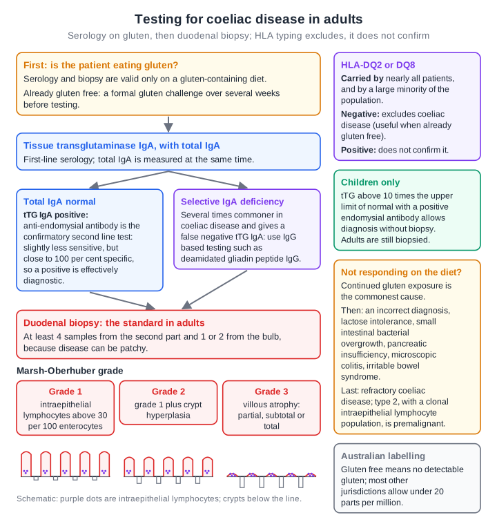
Flow chart for coeliac testing in adults. First, the patient must be eating gluten; if already gluten free, a formal gluten challenge over several weeks comes before testing. Tissue transglutaminase IgA is measured with total IgA. With normal IgA and a positive tTG IgA, anti-endomysial antibody is the confirmatory second line test, slightly less sensitive but close to 100 per cent specific. In selective IgA deficiency, several times commoner in coeliac disease and a cause of false negatives, IgG based testing such as deamidated gliadin peptide IgG is used. Both lead to duodenal biopsy, the standard in adults: at least 4 samples from the second part and 1 or 2 from the bulb. Marsh-Oberhuber grade 1 is intraepithelial lymphocytes above 30 per 100 enterocytes, grade 2 adds crypt hyperplasia and grade 3 is partial, subtotal or total villous atrophy, drawn as villi flattening and crypts deepening. Side panels: HLA-DQ2 or DQ8 is carried by nearly all patients and a large minority of the population, so a negative result excludes coeliac disease and a positive one does not confirm it; in children only, tTG above 10 times the upper limit with a positive endomysial antibody allows diagnosis without biopsy; non-response is most often continued gluten, then an incorrect diagnosis, lactose intolerance, bacterial overgrowth, pancreatic insufficiency, microscopic colitis or irritable bowel syndrome, and last refractory coeliac disease, whose type 2 is premalignant; Australian labelling requires no detectable gluten, where most other jurisdictions allow under 20 parts per million.
Physify teaching diagram (schematic). Tap the image to zoom.

From the lesson “Coeliac Disease, Villous Atrophy & Whipple Disease” in Coeliac & Small Bowel Disease
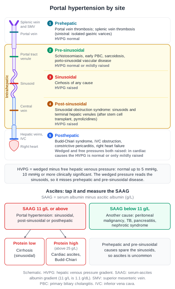
Portal hypertension by site, drawn along the path of blood from the splenic vein and superior mesenteric vein through the portal vein, the portal tract venule, the sinusoid and the central vein to the hepatic veins, inferior vena cava and right heart. 1, prehepatic: portal vein thrombosis, splenic vein thrombosis (sinistral portal hypertension with isolated gastric varices); HVPG normal. 2, intrahepatic pre-sinusoidal: schistosomiasis, early primary biliary cholangitis, sarcoidosis, porto-sinusoidal vascular disease; HVPG normal or mildly raised. 3, sinusoidal: cirrhosis of any cause; HVPG raised. 4, post-sinusoidal: sinusoidal obstruction syndrome, at the sinusoids and terminal hepatic venules, after stem cell transplantation or pyrrolizidine alkaloids; HVPG raised. 5, posthepatic: Budd-Chiari syndrome, IVC obstruction, constrictive pericarditis, right heart failure; wedged and free hepatic venous pressures both raised, so in cardiac causes the HVPG is normal or only mildly raised. HVPG is wedged minus free hepatic venous pressure, normal up to 5 mmHg and clinically significant at 10 mmHg or more; the wedged pressure reads the sinusoids, so it misses prehepatic and pre-sinusoidal disease. Ascites: the serum-ascites albumin gradient (SAAG, serum albumin minus ascitic albumin). A SAAG of 11 g/L or above (1.1 g/dL) means portal hypertension at or beyond the sinusoids; then a low ascitic protein suggests cirrhosis and a high protein, above 25 g/L, suggests cardiac ascites or Budd-Chiari syndrome. A SAAG below 11 g/L means another cause: peritoneal malignancy, tuberculosis, pancreatitis or nephrotic syndrome. Prehepatic and pre-sinusoidal causes spare the sinusoids, so ascites is uncommon.
Physify teaching diagram (schematic). Tap the image to zoom.

From the lesson “Staging Cirrhosis & Portal Hypertension” in Cirrhosis & Portal Hypertension
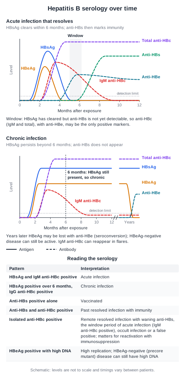
Two schematic graphs of hepatitis B markers against months after exposure, with antigens drawn as solid lines and antibodies dashed, and a dotted detection limit. Acute infection that resolves: HBsAg appears first and clears within 6 months; HBeAg rises and falls inside the HBsAg curve; IgM anti-HBc rises around the HBsAg peak and fades over the following months; total anti-HBc rises with it and persists; anti-HBe appears as HBeAg is lost; anti-HBs appears only after HBsAg has gone and persists, marking immunity. A shaded window lies between HBsAg clearing and anti-HBs becoming detectable, when anti-HBc (IgM and total), with anti-HBe, may be the only positive markers. Chronic infection: HBsAg persists beyond the 6 month mark, which defines chronic infection; HBeAg persists; IgM anti-HBc rises then falls to a low level; total anti-HBc persists; anti-HBs does not appear. After an axis break marked years, HBeAg may be lost with the appearance of anti-HBe (seroconversion); HBeAg-negative disease can still be active, and IgM anti-HBc can reappear in flares. A table reads the serology: HBsAg and IgM anti-HBc positive, acute infection; HBsAg positive over 6 months with IgG anti-HBc positive, chronic infection; anti-HBs positive alone, vaccinated; anti-HBs and anti-HBc positive, past resolved infection with immunity; isolated anti-HBc positive, remote resolved infection with waning anti-HBs, the window period of acute infection (IgM anti-HBc positive), occult infection or a false positive, which matters for reactivation with immunosuppression; HBeAg positive with high DNA, high replication, while HBeAg-negative (precore mutant) disease can still have high DNA.
Physify teaching diagram (schematic). Tap the image to zoom.

From the lesson “Hepatitis A and E, and Hepatitis B” in Viral Hepatitis

Free for Gastroenterology

Gastroenterology in the clinical exam

11 interactive short cases and 3 long cases for the RACP Divisional Clinical Exam: you examine, present and are marked on the five RACP domains.

See the clinical exam →

Open all of Gastroenterology

A membership opens every lesson and all 856 Gastroenterology questions, with the rest of the bank (11,500+ questions across 17 specialties), 15 timed mock exams, smart review and a daily plan to your exam.

The trial needs a card; cancel any time before it ends and you pay nothing. The free account needs no card.

Other specialties

Physify is a study aid for exam preparation only. It is not medical advice and must not be used to guide the care of any patient. Terms