RACP Divisional Written Exam · FRACP
Pulmonology for the written exam
Everything Physify has for Pulmonology: short lessons written to Australian guidance, exam-style questions with a worked explanation for each, and Bedside, our AI tutor, under every one. Try a question below, no sign-up.
- courses
- 24courses
- lessons
- 175lessons
- exam-style questions
- 661exam-style questions
- figures in the lessons
- 33figures in the lessons
Try one now · no sign-up
Pulmonology
Pick an answer to see the explanation
What's covered
24 courses, each a set of lessons with a quiz after them. The bar shows how many questions each has. Open a course to see its lesson outline.
- Australian Guidance Versus Overseas: Cardiovascular, Renal, Endocrine, Respiratory and Neurology4 lessons · 30 Qs
- Congenital Pulmonary Disorders11 lessons · 30 Qs
- Eosinophilic Disease7 lessons · 25 Qs
- Interstitial Lung Disease10 lessons · 30 Qs
- Landmark Trials 2016 to 2026: Critical Illness in Medical Patients5 lessons · 27 Qs
- Landmark Trials 2016 to 2026: Respiratory Medicine5 lessons · 27 Qs
- Landmark Trials: Endocrinology, Gastroenterology and Respiratory Medicine5 lessons · 30 Qs
- Lung Transplantation & Immunocompromised Hosts9 lessons · 21 Qs
- Miscellaneous Pulmonary Disorders12 lessons · 21 Qs
- Non-Cancer Pulmonary Masses8 lessons · 21 Qs
- Obstructive Airway Disease10 lessons · 30 Qs
- Occupational and Environmental Lung Disease5 lessons · 31 Qs
- Oxygen Therapy5 lessons · 28 Qs
- Pleural Diseases9 lessons · 41 Qs
- Pulmonary Diagnostics8 lessons · 25 Qs
- Pulmonary Hypertension4 lessons · 31 Qs
- Pulmonary Infections11 lessons · 31 Qs
- Pulmonary Physiology6 lessons · 26 Qs
- Pulmonary Rehabilitation & Preventive Medicine7 lessons · 25 Qs
- Pulmonary Vascular Disorders10 lessons · 31 Qs
- Re-emerging Respiratory Infections6 lessons · 25 Qs
- Respiratory Failure & Acute Respiratory Distress Syndrome8 lessons · 24 Qs
- Sarcoidosis5 lessons · 21 Qs
- Sleep-Disordered Breathing and Hypoventilation5 lessons · 30 Qs
From the lessons
33 figures sit in the Pulmonology lessons: ECGs, diagrams, films and clinical images, each with what to look for. Here are 4. Tap one to enlarge it.
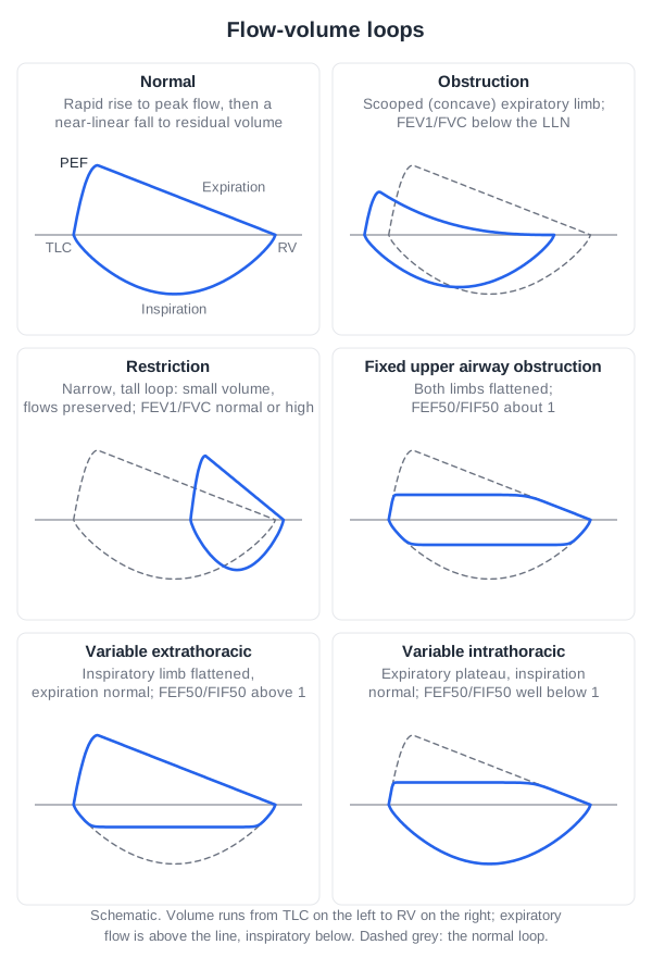
From the lesson “Lung Volumes, Diffusing Capacity and Flow-Volume Loops” in Pulmonary Diagnostics
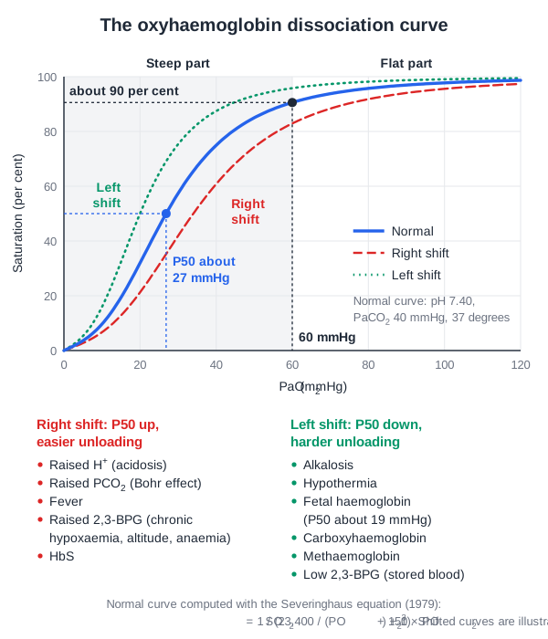
From the lesson “Oxygen Carriage, the Dissociation Curve and Dyshaemoglobins” in Pulmonary Physiology
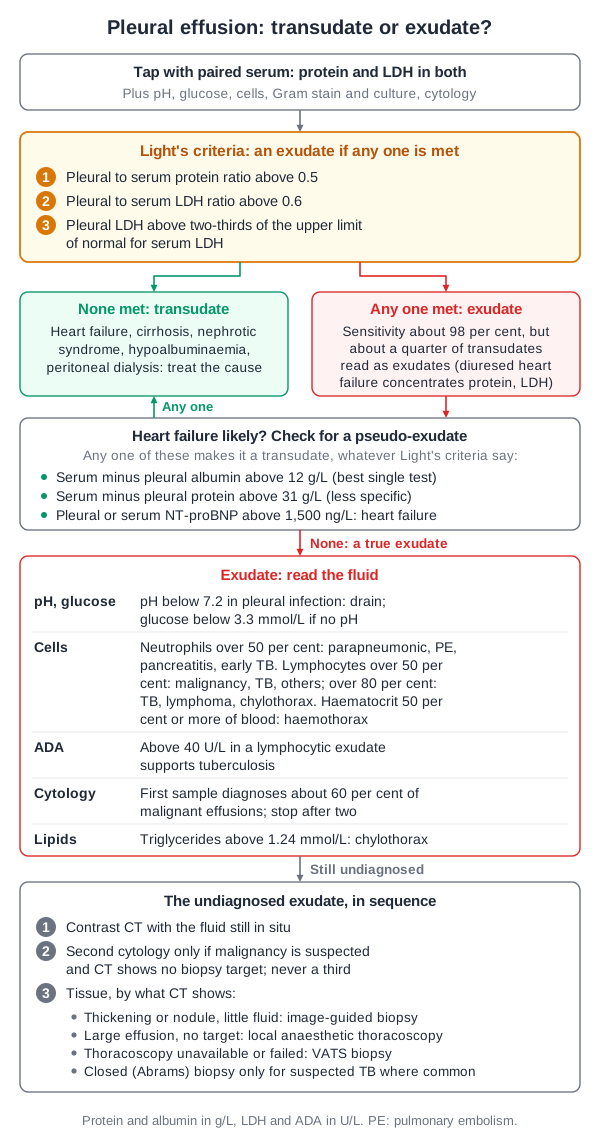
From the lesson “Pleural Effusion: Imaging, Sampling and Transudate versus Exudate” in Pleural Diseases
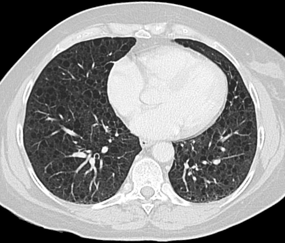
From the lesson “Lymphangioleiomyomatosis” in Interstitial Lung Disease
Free for Pulmonology
Pulmonology in the clinical exam
14 interactive short cases and 5 long cases for the RACP Divisional Clinical Exam: you examine, present and are marked on the five RACP domains.
See the clinical exam →Pulmonology on the blog
All posts →Alpha-1 Antitrypsin Deficiency: Trials of Note for the RACP Exam
Alpha-1 antitrypsin deficiency for the RACP exam: who to test (TSANZ 2020), levels and genotypes, augmentation from RAPID to Cochrane 2026, and fazirsiran.
Bronchiectasis: Trials of Note for the RACP Exam
Bronchiectasis for the RACP exam: the TSANZ 2023 work-up, 14-day antibiotics, macrolides after 3 exacerbations, inhaled antibiotics, and brensocatib (ASPEN).
Chronic Obstructive Pulmonary Disease: RACP Exam Notes
COPD for the RACP exam: the COPD-X stepwise approach, the 2024 Clinical Care Standard, SpO2 88 to 92 per cent, LTOT thresholds, and biologics (BOREAS, MATINEE).
Cystic Fibrosis: Key Points for the RACP Exam
Cystic fibrosis for the RACP exam: the 2023 Australian Standards of Care, sweat chloride cut-offs, annual review, and CFTR modulators from Trikafta to Alyftrek.
Idiopathic Pulmonary Fibrosis: Trials of Note for the RACP Exam
IPF for the RACP exam: TSANZ 2024 on antifibrotics at diagnosis, PBS nintedanib and pirfenidone, progressive pulmonary fibrosis, PANTHER, and FIBRONEER trials.
Pleural Infection and Empyema: Key Points for the RACP Exam
Pleural infection for the RACP exam: pleural fluid pH and LDH cut-offs, small-bore drains, tPA with DNase (MIST-2), Australian ADAPT dosing, and RAPID score.
Open all of Pulmonology
A membership opens every lesson and all 661 Pulmonology questions, with the rest of the bank (11,500+ questions across 17 specialties), 15 timed mock exams, smart review and a daily plan to your exam.
The trial needs a card; cancel any time before it ends and you pay nothing. The free account needs no card.