RACP Divisional Written Exam · FRACP
Neurology for the written exam
Everything Physify has for Neurology: short lessons written to Australian guidance, exam-style questions with a worked explanation for each, and Bedside, our AI tutor, under every one. Try a question below, no sign-up.
- courses
- 36courses
- lessons
- 302lessons
- exam-style questions
- 844exam-style questions
- figures in the lessons
- 28figures in the lessons
Try one now · no sign-up
Neurology
Pick an answer to see the explanation
What's covered
36 courses, each a set of lessons with a quiz after them. The bar shows how many questions each has. Open a course to see its lesson outline.
- Australian Guidance Versus Overseas: Cardiovascular, Renal, Endocrine, Respiratory and Neurology4 lessons · 30 Qs
- Bell's Palsy5 lessons · 25 Qs
- CIDP and Immune-Mediated Neuropathies9 lessons · 20 Qs
- CSF Pressure Disorders: Intracranial Hypertension and Hypotension7 lessons · 20 Qs
- Death by Neurological Criteria and Disorders of Consciousness5 lessons · 28 Qs
- Eagle Syndrome3 lessons · 6 Qs
- Epilepsy11 lessons · 31 Qs
- Functional Neurological Disorder7 lessons · 25 Qs
- Guillain-Barre Syndrome6 lessons · 20 Qs
- Head Trauma5 lessons · 6 Qs
- Hereditary and Acquired Ataxias9 lessons · 20 Qs
- Hereditary Spastic Paraplegia5 lessons · 6 Qs
- Hydrocephalus6 lessons · 6 Qs
- Infections of the Central Nervous System10 lessons · 21 Qs
- Landmark Trials 2016 to 2026: Neurology and Stroke5 lessons · 27 Qs
- Landmark Trials: Neurology, Rheumatology, Geriatrics and Acute Care5 lessons · 29 Qs
- Motor Neurone Disease9 lessons · 30 Qs
- Movement Disorders, Parkinson's Disease & Parkinson's Plus Syndromes20 lessons · 30 Qs
- Multiple Sclerosis14 lessons · 30 Qs
- Muscular Dystrophy & Myopathy14 lessons · 30 Qs
- Neuro-Ophthalmological Disorders11 lessons · 31 Qs
- Neurocutaneous Syndromes7 lessons · 26 Qs
- Neurological Diagnostics8 lessons · 25 Qs
- Neurological Emergencies8 lessons · 27 Qs
- Neuromuscular Junction Disorders12 lessons · 26 Qs
- Neurophysiology: Motor Circuits, Pain, Sleep and CSF8 lessons · 30 Qs
- Neurotransmitters and Receptor Signalling6 lessons · 30 Qs
- Peripheral Neuropathy8 lessons · 30 Qs
- Primary Headache Disorders9 lessons · 30 Qs
- Secondary Headache Disorders8 lessons · 25 Qs
- Sleep Disorders in Neurology9 lessons · 20 Qs
- Spinal Anatomy & Pathology18 lessons · 36 Qs
- Spinocerebellar Ataxia4 lessons · 6 Qs
- Stroke, Transient Ischaemic Attack and Common Causes17 lessons · 31 Qs
- Subclavian Steal Syndrome3 lessons · 6 Qs
- Vertigo7 lessons · 25 Qs
From the lessons
28 figures sit in the Neurology lessons: ECGs, diagrams, films and clinical images, each with what to look for. Here are 4. Tap one to enlarge it.
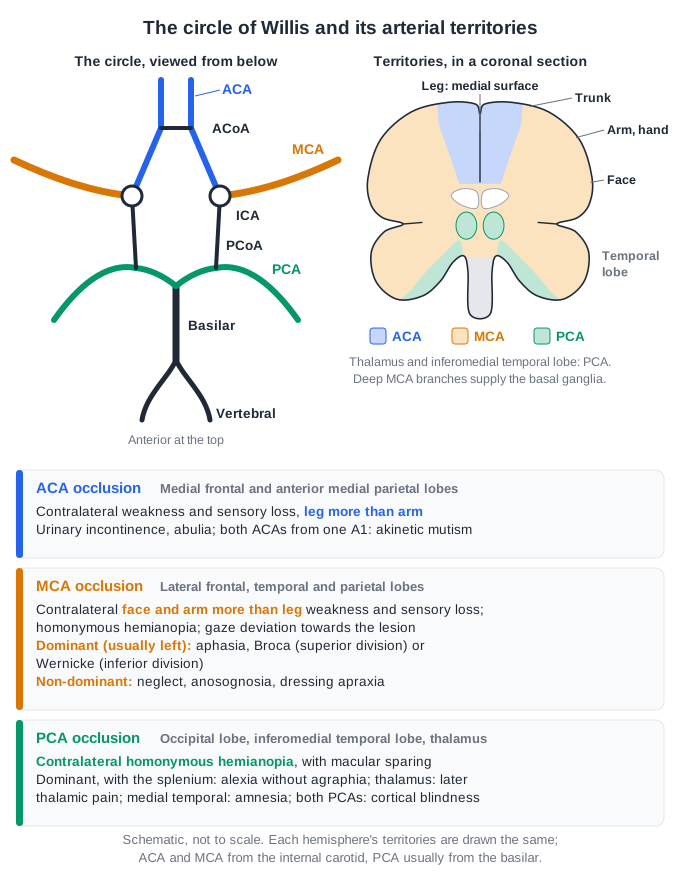
From the lesson “Vascular Territories, Lacunar Syndromes and Localisation” in Stroke, Transient Ischaemic Attack and Common Causes
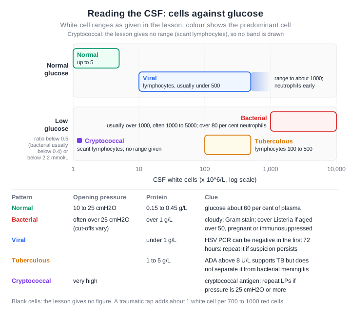
From the lesson “Bacterial Meningitis: Organisms, Clinical Features and the CSF” in Infections of the Central Nervous System
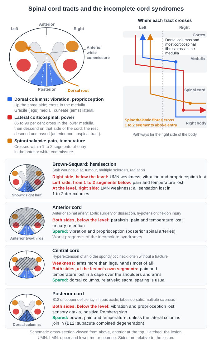
From the lesson “Incomplete Cord Syndromes: Central, Brown-Sequard, Anterior and Posterior” in Spinal Anatomy & Pathology
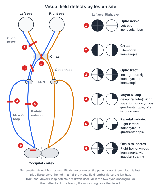
From the lesson “Retrochiasmal Field Loss, Hemianopia and Charles Bonnet Syndrome” in Neuro-Ophthalmological Disorders
Free for Neurology
Neurology in the clinical exam
29 interactive short cases and 7 long cases for the RACP Divisional Clinical Exam: you examine, present and are marked on the five RACP domains.
See the clinical exam →Neurology on the blog
All posts →Emerging Therapies and Interventions in Epilepsy: RACP Exam Notes
Epilepsy for the RACP exam: SANAD II first-drug choice, Australian valproate advice, ESETT for status, Austroads driving rules and cenobamate and azetukalner.
Migraine: Emerging Therapies and Innovations
Migraine for the RACP exam: CGRP antibodies and Botox on the PBS, erenumab off it, gepants on private script, the PACAP antibody, and safety in pregnancy.
Diagnostic and Therapeutic Advances in CNS Infections: RACP Exam Notes
CNS infections for the RACP exam: meningitis and dexamethasone, the ANZ encephalitis guideline, Japanese encephalitis in Australia, mNGS and brain abscess.
Emerging Therapies and Innovations in Neuro-Ophthalmology: RACP Exam Notes
Neuro-ophthalmology for the RACP exam: optic neuritis and MOGAD, tocilizumab for GCA and teprotumumab for thyroid eye disease on the PBS, and LHON therapies.
Emerging Therapies for Guillain-Barré Syndrome: RACP Exam Notes
Guillain-Barré syndrome for the RACP exam: IVIg under the National Blood Authority criteria, EAN/PNS 2023, no second IVIg dose, eculizumab and C1q inhibition.
Emerging Therapies for Huntington's Disease: RACP Exam Notes
Huntington's disease for the RACP exam: tetrabenazine on the PBS, valbenazine in KINECT-HD, tominersen stopped, PROOF-HD negative, AMT-130's 3-year results.
Open all of Neurology
A membership opens every lesson and all 844 Neurology questions, with the rest of the bank (11,500+ questions across 17 specialties), 15 timed mock exams, smart review and a daily plan to your exam.
The trial needs a card; cancel any time before it ends and you pay nothing. The free account needs no card.